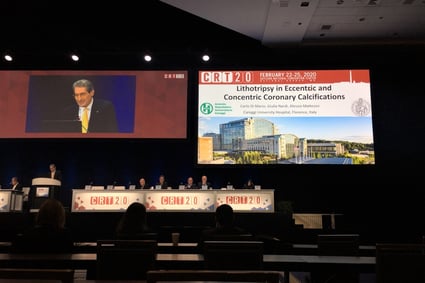
Q&A: Imaging with IVL in Concentric & Eccentric Ca2+
Prof. Carlo Di Mario, Director of Structural Interventional Cardiology at the Careggi University Hospital in Florence, Italy, and other cardiologists in Florence recently published their initial real-world experience when using intravascular imaging while treating coronary lesions with Intravascular Lithotripsy (IVL).
The publication in Cardiovascular Revascularization Medicine, entitled “Intravascular imaging to guide lithotripsy in concentric and eccentric calcific coronary lesions,” showed similar positive outcomes in more complex real-world patients who failed other calcium modification strategies before turning to imaging-guided IVL, compared to the patients treated with de novo lesions in DISRUPT CAD I & II. What’s more, his analysis also looked at the arc of calcium to see if there were any differences in outcomes. His results, in his own words, were “unexpected.”
We had the privilege of connecting with Prof. Di Mario to learn more about the publication, its key takeaways and some of his practical recommendations as one of the earliest users of Shockwave IVL in the coronaries.
Read the publication and enjoy his Q&A:
Question: What are the current challenges in treating calcified eccentric lesions and solely using angiography to guide IVL delivery?
Prof. Di Mario: The main issue is that we have no certainty, only based on angiography, of the true extension of the calcium arc. Thinner superficial calcium sheets can be less visible than the contralateral thicker layers giving the false impression of an eccentric calcification and vice versa parallel rails of calcium can be present in one view even below the 180 degree arc. This is the main limitation of the manuscript that Nef and colleagues recently submitted to Circulation Interventions on behalf of all the DISRUPT I and II Investigators. The distinction was only based on the careful angiographic review done at Yale by the Angiographic Core Lab of Dr Alexandra Lansky which overcomes the subjectivity of the Investigators but cannot eliminate the inherent limitations of angiography, known since the seminal IVUS paper of Mintz et al in the late nineties.
Question: What were your study’s goals – what did you seek to understand going into the research?

Prof. Di Mario: Our study is much smaller than Nef’s combined analysis of 180 patients with only 31 lesions in 28 patients but they came not from the strict adherence to the DISRUPT trial protocol but from the real world, with patients selected based on severe calcium load confirmed with IVUS/OCT, patients that failed predilatation, patients with Rotablator used to cross the lesion initially but still with an incomplete final lesion expansion. More importantly, in all these patients the distinction between eccentric and concentric lesions was based on the gold standard, IVUS or OCT.
Question: What were your study’s key findings?
Prof. Di Mario: Lesion minimal lumen area measured with OCT/IVUS was 7.06 mm2 in the eccentric lesion group and 7.13 mm2 in the concentric lesion group.
Question: Why was the similar stent expansion and acute gain observed in both concentric and eccentric calcified lesions an unexpected outcome?
Prof. Di Mario: We expected that concentric lesions had greater potential benefit from IVL and some expert users were even supporting the idea that, in order to select these lesions appropriately, IVUS/OCT was indispensable before IVL. On the contrary, we had similar findings to the angiographic analysis from DISRUPT I and II where residual stenosis >30% was less than 3% with QCA both in eccentric and concentric lesions. This suggests that the pragmatic approach proposed in these trials with angiography only to select patients for IVL obtains excellent outcome without complications in both types of lesions.
Question: How does the use of imaging change the way that someone uses IVL?
Prof. Di Mario: I think the main difference is the ability to avoid risky high pressure dilatations with balloon dogboning and possible vessel rupture in lesions meeting the classical Fujino OCT criteria or anyway with >180 degrees and long calcifications with IVUS.
Question: From your experience, are there certain lesions characteristics or anatomical locations that most often benefit from imaging-guided IVL?
Prof. Di Mario: Long lesions, especially in tapering vessels such as the proximal-mid LAD, may require more than one IVL balloon, both because multiple activations are needed along the vessels and because the more proximal balloon diameter should be ½-1 mm greater than the diameter of the distal balloon. IVUS/OCT are ideal for these complex lesions and very helpful, especially OCT, to confirm effective calcium cracking and optimal final stent expansion and strut apposition.
Question: How would you counsel someone without access to imaging to optimize their use of IVL?
Prof. Di Mario: Even in expert imaging centers there would be instances when this is difficult to use, like in primary PCI in the middle of the night or due to cost or organizational difficulties. Long rails of calcium in proximal large vessels were the chief inclusion criterion in the DISRUPT trials. Wait a second before injecting contrast to examine the running images of the vessel without contrast in various views. As the angiographic DISRUPT subanalysis showed and our imaging study confirmed all the lesions qualifying angiographically have a benefit from IVL both in terms of optimal expansion and reduction of complications.
Important Safety Information - Coronary
Caution: In the United States, Shockwave C2 Coronary IVL catheters are investigational devices, limited by United States law to investigational use. DISRUPT CAD III Study
Shockwave C2 Coronary IVL catheters are commercially available in certain countries outside the U.S. Please contact your local Shockwave representative for specific country availability. The Shockwave C2 Coronary IVL catheters are indicated for lithotripsy-enhanced, low-pressure balloon dilatation of calcified, stenotic de novo coronary arteries prior to stenting. For the full IFU containing important safety information please visit: https://shockwavemedical.com/clinicians/international/coronary/shockwave-c2/


